|
|
|
|
|
|
|
|
|
ménisque Each knee has two menisci, the so-called medial meniscus on the inner part of the knee and the lateral meniscus on the outside of the knee. The menisci are considered to be a form of cartilage (therefore the term "torn cartilage" when a meniscus is torn). The menisci are located between the femur (thigh bone) and the tibia (shin bone). As seen in the accompanying figures, the menisci cannot completely cover the tibia, thus leaving an area of direct contact between the cartilage of the femur and that of the tibia. The menisci contribute to the stability of the knee (minimizing abnormal movements) and also act as a shock absorber. A tear of the meniscus may not compromise the stability of the knee if the ligaments are intact. However, a large tear can predispose to arthritis, especially if a large portion of the meniscus is surgically removed. |
|
|

|
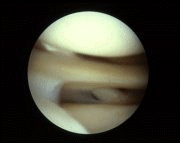
The lateral meniscus is located between the femur (on top in the picture) and the tibia (below in the picture). Both bones are covered with cartilage which appears white or beige in the picture. The rope-like structure seen going from top left to bottom right behind the meniscus, is the tendon of the popliteus muscle. ménisq |
|
Arthroscopic view of the lateral meniscus |
|
ménisque
Meniscal tears are divided into traumatic and non-traumatic tears : Traumatic tears can occur in knees that are essentially stable or unstable. Non-traumatic (atraumatic) lesions are divided into degenerative ("wear and tear") and tears seen in conjunction with arthritis.
|
|
Meniscal tears can sometimes be found in conjunction with arthritis (wearing out of the articular cartilage which lines the end of the femur and the tibia). The arthritis in itself can be a source of pain and removal of the torn cartilage may not improve the patient’s symptoms in this particular setting. Certainly, plain x-rays (taken in the "Schuss" position), as well as direct visualization of the knee during an arthroscopic procedure will help detect these arthritic conditions.
|
|
Benign neglect. In certain conditions it is possible to leave the meniscus alone. For example, if the tear is small and the surgeon does not feel that it is a significant source of pain, it may be left alone. |
|
Repair. In rare cases, the cartilage can be stitched back together. The knee must be stable for this repair to have a chance of working. |
|
|
"Meniscectomy." This involves the removal of the portion of meniscus which is torn. |
There is no medical need for surgery in the setting of a torn meniscus. Indeed the only reason for recommending surgery is for pain relief. It is up to the patient to decide whether he or she is troubled enough by the torn meniscus to warrant a surgical procedure. It is not for the surgeon to determine whether a patient needs surgery. The surgeon must, however, give the patient all the information that he or she needs to make an informed decision. There is no harm in leaving a torn meniscus in place. A delay in surgery will not make the surgery more difficult nor will it be harmful to the patient in any way. The only downside to waiting is naturally the persistence of pain and possible disability as the patient waits for surgery. Surgery should not be carried out if the pain is minimal and without effect on activities of daily living. In such mild cases, a non-operative regimen is usually successful.
On the other hand, if symptoms are significant, it is a shame not to take advantage of a relatively simple procedure which can essentially eliminate the pain and disability.